Thursday, December 29, 2016

How Absorption Works

By Jonathan Alfred

All those food we eat don’t just turn into poop straight away. The body actually absorbs it at different points throughout the GIT system. Now lets follow this wonderful roller coaster like journey that food goes through during absorption.
Now that's one hell of a ride

Absorption Process
What is it?
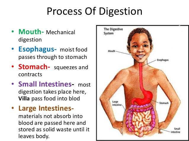
To talk about absorption we have to give the small intestine a not so small introduction. The small intestine is the part of the gastrointestinal tract between the stomach and the large intestine, and is where most of the end absorption of food takes place. The small intestine has three distinct regions which are the duodenum, jejunum, and ileum. The duodenum receives bile and pancreatic juice through the pancreatic duct, controlled by the sphincter of Oddi. The primary function of the small intestine is the absorption of nutrients and minerals from food.

How it works?
Digested food is now able to pass into the blood vessels in the wall of the intestine through either diffusion or active transport. The small intestine is the site where most of the nutrients from ingested food are absorbed. The inner wall, or mucosa, of the small intestine is lined with simple columnar epithelial tissue. Structurally, the mucosa is covered in wrinkles or folds called plicae circulares, which are considered permanent features in the wall of the organ. They are distinct from rugae which are considered non-permanent or temporary allowing for distention and contraction. From the plicae circulares project microscopic finger-like pieces of tissue called villi (Latin for "shaggy hair"). The individual epithelial cells also have finger-like projections known as microvilli. The functions of the plicae circulares, the villi, and the microvilli are to increase the amount of surface area available for the absorption of nutrients, and to limit the loss of said nutrients to intestinal fauna.

Each villus has a network of capillaries and fine lymphatic vessels called lacteals close to its surface. The epithelial cells of the villi transport nutrients from the lumen of the intestine into these capillaries (amino acids and carbohydrates) and lacteals (lipids). The absorbed substances are transported via the blood vessels to different organs of the body where they are used to build complex substances such as the proteins required by our body. The material that remains undigested and unabsorbed passes into the large intestine.

Absorption of the majority of nutrients takes place in the jejunum, with the following notable exceptions:
Iron is absorbed in the duodenum.
Vitamin B12 and bile salts are absorbed in the terminal ileum.
Water and lipids are absorbed by passive diffusion throughout the small intestine.
Sodium bicarbonate is absorbed by active transport and glucose and amino acid co-transport.
Fructose is absorbed by facilitated diffusion.

And this is where and how the magic happens

Saturday, December 17, 2016

Digestion process


DIGESTION PROCESS
by Nur Annissa

  • MOUTH
Digestion starts when food is ingested at the mouth. Chewing breaks down the food
into small pieces so that it is easily digested. Saliva breaks down the food for the digestive system to absorb and use.


  • ESOPHAGUS
The esophagus is a long, muscular tube that connects the throat to the stomach. Food is pass to the stomach by a process called peristalsis. Peristalsis is a series of contraction of muscle along the esophagus for the food ball. At the esophagus also is a sphincter which is called lower esophageal sphincter. the function of this sphincter is to prevent backwards movement of the food.


  • STOMACH
The stomach secretes acid and enzymes that will further break down food.


  • SMALL INTESTINE
The small intestine is made up of segments. these segments are the duodenum, jejunum, and ileum. The duodenum continues to break down food while the jejunum and ileum absorbs the nutrients into the bloodstream.


  • LARGE INTESTINE
The large intestine is also known as the colon. Waste products from the digestion process is passed through here by the process peristalsis. when the colon becomes full of waste products, it empties its contenys into the rectum for elimination.


  • RECTUM
The rectum connects the colon to the anus. The function of the rectum is to receive stool from the colon, let the brain know that there's stool to be eliminated, and holds the stool until elimination. here, there's a sphincter that controls the opening to the anus.

Tuesday, December 13, 2016

Defecation Process

Defecation

Below is the process of defecation which is i included the detail information about it. Have you wonder how does the procedure is ?


Well, this is the answer to all of your mystery !


Fecal continence is maintained by two sphincters that surround the anal canal. The internal anal sphincter is an extension of the muscularis externa, and thus is smooth muscle innervated by enteric neurons found in the myenteric plexus. 


The external anal sphincter is skeletal musclethat can be consciously controlled, and is innervated by somatic efferent neurons. The figure is a schematic sagittal view of the lower rectum and anus showing the anal sphincters.

Another skeletal muscle that contributes to the maintenance of fecal continence is the puborectalismuscle. This muscle loops around the rectum like a sling, pulling it forward to create a more acute angle between the rectum and the anal canal (the anorectal angle). 

During defecation, there is conscious relaxation of the puborectalis muscle, which creates a more open anorectal angle and allows for a straigther passage through the anal canal. Look at Figure 1 in this paper for an illustration of the role of the puborectalis muscle.


This is our face when we are doing defecation process 

Monday, December 5, 2016

TIPS TO HAVE A HEALTHY DIGESTIVE SYSTEM

TIPS ON HEALTHY DIGESTIVE SYSTEM
by Nur Annissa


1. Make sure that your diet consists of fibre.

Food that are rich in fibre helps the peristalsis process by ensuring it goes well. this will prevent constipation and diarrhea.


2. avoid fats
i always tell my friends to avoid any sugary food or drinks. i do this because i stay true to the saying "too much of anything will make you sick".

this is because you'll need some fat in your diet BUT too much fat in your diet will cause harm to your body. Fats can also cause you to be- well, fat.


so by avoiding food that have fats in it, you can also maintain your body shape. fats can also cause constipation as it slows down the process of peristalsis.


3. Drink water.
i cannot stress enough how drinking enough water can change your life. Our body NEEDS water to function properly. Water helps our body to eliminate waste easily, in other words, water helps the process of defecation and urination.


4. Avoid sodas.
I can proudly say i've been clean from soda for nearly a year now. I used to love to drink coke, sprite and all sodas, but after realizing how bad is it for my health, i stopped. sodas are full of acids and chemicals that can harm the natural acids inside our digestive system.


5. Exercise.
the easiest way to keep our digestive system (and also our body) healthy is to exercise. you don't have to go to the gym to exercise, just simple brisk walking will work.

this will allow blood to circulate properly from the heart to the digestive system. exercises like sit ups and crunches also helps to strengthen the abdominal muscle.
this will cause the small intestine to function properly because the muscles are consistently moving.


6. eat on schedule.
eating at the right time everyday can do wonders to your digestive system. by following a schedule, you can avoid gastritis.


REFERENCES

  • Powerful Tips to Improve Your Digestive System's Health: From a Conversation with Nancy Spahr, CBE & Colon Therapist by Nancy Spahr; retrieved from http://bodyecology.com/articles/nancy_spahr_colon_therapist.php 
  • 5 Tips For Keeping Your Digestive System Healthy by Dr. Edward Group DC, NP, DACBN, DCBCN, DABFM; Published on http://www.globalhealingcenter.com/natural-health/how-to-keep-your-digestive-system-healthy/

Sunday, December 4, 2016

Anatomy and Physiology of the Digestive System

Overview of the Digestive System

By Zaki
So, what in the world is the Digestive System? Well, the Digestive System is composed of two groups of organ, which are the the Gastrointestinal Tract (GI) and the Accessory Digestive organs. The Gastrointestinal Tract (GI) is a continuous tube that extends from the mouth to the anus through thoracic and abdominopelvic cavities.
From the back.
From the front.
What organs include in the Gastrointestinal Tract? The organs of the Gastrointestinal Tract include the mouth, most of the pharynx, esophagus, stomach, small intestine, and large intestine.

And What organs include in the Accessory Digestive? you might ask, it include the teeth, tongue, salivary glands, liver, gallbladder, and pancreas. The teeth aid in the physical breakdown of food, and the tongue assists in chewing and swallowing. The other accessory digestive organs, however, never come into direct contact with food. They mainly produce or store secretions that flow into the GI tract through ducts. Basically the secretions aid in the chemical breakdown of food.

What else should you know about the Digestive System? There's one thing that you should know and that is the length of the Gastrointestinal Tract which is about 5-7 meters. THAT LONG! Incase you don't have a rope, you know what to do right? Well yeah buy a rope! 


Functions of the Digestive System


Overall, the digestive system performs six basic processes:


  1. Ingestion: Taking food into mouth.
  2. Secretion: Release of water, acid, buffers, and enzymes into lumen of GI tract.
  3. Mixing and Propulsion: Churning and Propulsion of food through GI tract.
  4. Digestion: Mechanical and Chemical breakdown of food.
  5. Absorption: Passage of digested products from GI tract into blood and lymph.
  6. Defecation:  Elimination of feces from GI tract.


The flow of taking food until you poop!


Layers of the Gastrointestinal Tract 


Before we go deeply on the anatomy and physiology, let me educate you guys the layers of the GI tract. The wall of GI tract from the lower esophagus to the anal has the same basic, four-layered arrangement of tissues. The four layers of the tract, from deep to superficial, are the Mucosa, Submucosa, Muscularis, and Serosa or Adventitia (basically the same). So yeah, that's it! Only 4 layers guys! It's that easy. Let me show you guys the structure of the layers below.

The 4 layers of the Gastrointestinal Tract.

What is Peritoneum?


The peritoneum is the largest serous membrane of the body, it consists of a layer of simple squamous epithelium with an underlying supporting layer of areolar connective tissue. The peritoneum is divided into the Parietal Peritoneum, which lines the wall of the abdominopelvic cavity, and the Visceral Peritoneum, which covers some of the organs in the cavity and is their serosa.
The slim space containing lubricating serous fluid that is between the parietal and visceral portions of the peritoneum is called the Peritoneal Cavity. Unlike the pericardium and pleurae, which smoothly cover the heart and lungs, the peritoneum contains large folds that weave between the viscera. The folds bind the organs the one another and to the walls of the abdominal cavity. They also contain blood vessels, lymphatic vessels, and nerves that supply the abdominal organs. There are five major peritoneal folds which are the greater omentum, falciform ligament, lesser omentum, mesentry, and mesocolon.


Midsagittal section showing the peritoneal folds.


The Gastrointestinal Tract Organs


Mouth 


In human anatomy, the mouth is the first portion of the alimentary canal that receives food and saliva. The primary role of a mouth its obviously to eat, but other than eating, the mouth also plays a good role in gossiping (I mean communication). The mouth is normally moist, because it is lined with a mucous membrane, and contains the teeth to chew your food. The lips mark transition from mucous membrane to skin, which covers most of the body. 

The mouth
 

Pharynx


The pharynx is actually part of the throat that is behind the mouth and nasal cavity and above the oesphagus and the larynx, or any tubes going down to the stomach and the lungs. The pharynx is an organ which can be found in the vertebrates and invertebrates, though the structure is not universally the same across all of those species obviously. Pharynx is part of the digestive system and also of the conducting zone of the respiratory system. Our pharynx is actually divided into three sections which are the nasopharynx, the oropharynx and laryngopharynx. Well, if you really want to know what these three sections is all about, you can go for a book.


The pharynx with the three sections.


Esophagus


The esophagus is mainly called the "tube of food". I just created that. It's an organ in vertebrates through which food passes from the pharynx to the stomach. The esophagus is a fibromuscular tube, about 25 damn centimetres in adult. It travels behind the trachea and heart, passes through the diaphragm and empties into the uppermost region of stomach. If you just imagine the tube is something like the water slides.

Imagine these fellas are the food you ate just now and they are traveling down to your stomach.

The esophagus.
The esophagus secretes mucus and transport food into the stomach. It does not produce digestive enzymes, and it does not carry on absorption.


Stomach


The stomach is probably the happiest part you tap after you eat something. Everyone does that! It is involved in the second phase of digestion, following chewing. The stomach is located between the esophagus and the small intestine. It secretes digestive enzymes and gastric acid to aid in food digestion. The pyloric sphincter controls the passage of partially digested food from the stomach into the duodenum where peristalsis takes over to move this through the rest of the intestines. You can see the pyloric sphincter in the diagram below.

The stomach in you.


The functions of the stomach are:
  1. Mixes saliva, food, and gastric juice to form chyme.
  2. Serves as reservoir for food before release into small intestine.
  3. Secretes gastric juice, which contains HCl (kills bacteria and denatures protein), pepsin (begins the digestion of proteins), intrinsic factors (aids absorption of vitamin B12), and gastric lipase (aids digestion of triglycerides).
  4. Secretes gastrin into blood.


Small Intestine


The small intestine is between the stomach and the large intestine, and is where most of the end absorption of food takes place. The small intestine actually has three regions and there are the duodenum, jejunum, and ileum. The primary function of small intestine is to absorp nutrients and minerals from food. Easy work there right?

The small intestine with three different regions.

The functions of the small intestine:
  1. Segmentations mix chyme with digestive juices and bring food into contact with mucosa for absorption, peritalsis propels chyme through small intestine.
  2. Completes digestion of carbohydrates, proteins, and lipids, begins and completes digestion of nucleic acids.
  3. Absorbs about 90% of nutrients and water that pass through digestive system.


Large Intestine


The large intestine is the last part of GIT system. Water is absorbed here and the remaining waste material is stored as poop before being removed by pooping. Large intestine is the combination of the cecum, colon, rectum, and also anal canal.
The large intestine with its combination.

The functions of the large intestine:
  1. Haustral churning, peritalsis, and mass peristalsis drive contents of colon into rectum.
  2. Bacteria in large intestine convert proteins to amino acids, break down amino acids, and produce some B vitamins and vitamin K.
  3. Absorbing some water, ions, and vitamins,
  4. Forming feces.
  5. Defecating (emptying rectum).


The Accessory Digestive Organs


Teeth


The teeth is functioned to mechanically break down food by cutting and crushing them in preparation before you swallow and for further digestion. Without teeth, you'll be having soups for your entire life.

That's you having soup with no teeth.
 Just for your information, there are four types of teeth and there are incisors, canines, premolars and molars each with specific function.

 
Thats my teeth right there!

Tongue

The tongue is a muscular organ in the mouth and it manipulates food for chewing, and is used in the act of swallowing. Without tongue, you can't eat and also talk and also to taste. It is senstive and kept moist by the saliva. Just for your information, the tongue serves as a natural means of cleaning the teeth.

The tongue
 
Liver and Gallbladder

The liver is a vital organ. In human, it is located in the upper right quadrant of the abdomen, below the diaphragm. The liver has a wide range of functions, including detoxification of various metabolites, protein synthesis, and the production of biochemicals necessary for digestion. It produces bile, an alkaline compound which aids in digestion via the emulsification of lipids. The gallbladder, a small pouch that sits just under the liver, stores bile produced by the liver.

Shows the liver in the human body.

The liver.

 The gallbladder is a small organ where bile is stored and concentrated before it is released into the small intestine. Just so you know, humans can live without a gallbladder. It sits just beneath the right lobe of the liver.

The gallbladder.


Pancreas


The pancreas is a glandular organ in the digestive system and also endocrine system. It is located in the abdominal cavity behind the stomach. It secretes pancreatic juice containing digestive enzymes that assist digestion and absorption of nutrients in the small intestine. These enzymes help to further break down the carbohydrates, proteins, and lipids in the chyme.

The pancreas.
 
Well that's all folks. Thank god I helped you with some educational info right? You're welcome.


Reference


  1. Principles of Anatomy and Physiology, Volume 2, 13th Edition by Gerard J. Tortora and Bryan Derrickson.


Wednesday, November 30, 2016

A Brief Introduction...

Hi there! Welcome to our blog!

The purpose of this blog is to educate the public (you) about the ingenious ways that our bodies go through to carefully manage all the things that we stuff our face with every. single. day!

It's a daunting task but never fear! Our bodies are well equipped with different systems and organizations to deal with it all. So, come what may, everything that we eat, be it good or bad, will eventually find its way out of us.

We've specifically designed our posts to be as reader-friendly as possible (read; we'll use as little medical jargon as we can, we promise!) so that educating yourselves through our posts will be as smooth as butter spreading across a piece of warm toast! Mmm, yummy...

Our topics will include;


  1. The Parts and The Functions of a Normal Digestive System (Anatomy & Physiology)
  2. Chewing!
  3. Swallowing!
  4. Digesting!
  5. Absorption...?
  6. Pooping!
  7. Tips to maintain a healthy digestive system
  8. Diseases of The Digestive System

Stay tuned!

Tuesday, November 29, 2016

Chewing!

by Afrina Ariff

So I thought it would be a good idea to write this out while eating. As you are reading this, I am about to dive into a pack of Chicken-flavoured Mamee Monster. Haven't had this in a very long time. Probably not the best thing to have at this hour but I am willing to sacrifice my able-bodied self for science!!!

*opens pack of Mamee*

Right, let's be honest with ourselves, have we ever given thought to what happens when we stick a piece of food in our mouth? How it is able to magically travel down our throats and fill up our stomachs to satisfy our insatiable and perpetual hunger? No? Understandable. 
The ~explosion~ of flavours usually distracts us. (This Mamee however, man is it salty...)

Before food can be swallowed, obviously, we gotta chew it first. Aaaaaaaand how do we chew food?
*WITH OUR TEETH!!!* Well done kids!

Image result for person with no teeth
Except for this guy. But don't feel bad for him tho. He doesn't seem to be bothered by his predicament.


In all seriousness, food needs to be chewed before it can be swallowed. If not you'll choke. and probably die if no one around you knows what to do about it. (psst! we suggest reading up about the Heimlich Maneuver.)


Chewing, or it's fancy scientific term, Mastication, is a process where food is broken down into small pieces with the help of the teeth and tongue. As you may already know, we've got different types of teeth to get the job done;


Related image

As you can see from the infographic above, us humans 4 types of teeth, each with its own unique function.

It makes perfect sense that the pointier teeth such as the Canines, Premolars and Incisors, are located nearest to the entrance of the mouth, because that's where the food we're about to stuff our face with comes in contact first.


Image result for person biting into food
Exhibit A : This woman's Incisors, Canines and Premolars are in full view, out and ready to bite into the piece of meat. She likes her meat rare by the looks of it.


It also makes perfect sense that the Molars, responsible for grinding up food after it has been bitten off, is located at the back and sides of the mouth, preparing the ground up food that has been mixed together with saliva, now called a bolus, to be swallowed.

Now the teeth don't do all the overwhelming work by themselves. They go hand in hand with specific muscles to produce specialized movements. These group of muscles are called the Muscles of Mastication. Let's go through them one by one;

1. Temporalis (tem-pow-ral-lisMuscle
 Image result for temporalis muscle

The temporalis muscle, located at the side of the head, is responsible to lift the jaw to bite into and CRUSH food between the Molars.

2. Masseter (ma-seh-turh) Muscle

Image result for masseter muscle

This muscle, which is located at the jaw, also plays the same role as the Temporalis muscle which is to lift the jaw when biting into food. It also enables us to clench our teeth in a fiery fit of rage when the waiter messes up our order.

3. Medial and Lateral Pterygoid (terry-goid)

Image result for medial pterygoid muscle function

The Pterygoids can be considered as the "connector" of the first two muscles mentioned. They allow our jaw to open and close and move from side to side.

To summarize, I've created a scenario and put all these pieces of information together in an orderly sequence;

1. You smell food. You want to eat food.

2. You find food (the last piece of chicken from last night's roast - a drumstick).

Related image
oh man

3. You think, there's no time for cutlery and plates, this needs to be IN me now.

4. You open your mouth (with the help of the Medial and Lateral Pterygoids) and bite into the chicken (using your Incisors if you're biting from the front of the mouth, the Canines and the Premolars if you're biting from the side).

5. You chew. (up and down movement of the jaw provided by all the muscles of mastication; grinding of the piece of food by the Molars).

6. When you're happy with the sludge that you've produced (formation of Bolus) it's finally time to SWALLOW.

Swallowing is another process that will be explained in the next post.

Until then, this has been Afrina and now you know how Chewing works! Tata!


Delectable!


REFERENCES

1.     Tortora, Gerard J., et al, “Organization, Support and Movement, and Control Systems of the Human Body 13th Edition”, John Wiley & Sons (Asia) Pte. Ltd., 2011.